<--- Back to Details
First PageDocument Content
Brain / Cerebrum / Neuroscience / Nervous system / Neuroanatomy / Visual system / Central nervous system / Visual cortex / Retinotopy / Occipital lobe / Cerebral cortex / Calcarine sulcus
Date: 2013-01-03 08:42:06
Brain
Cerebrum
Neuroscience
Nervous system
Neuroanatomy
Visual system
Central nervous system
Visual cortex
Retinotopy
Occipital lobe
Cerebral cortex
Calcarine sulcus

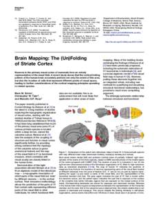
Brain Mapping: The (Un)Folding of Striate Cortex

Add to Reading List

Source URL: color.psych.upenn.edu

Download Document from Source Website

File Size: 222,04 KB

Share Document on Facebook

Similar Documents

Large Animal Miniboard ExamWith Borna disease in horses, pathognomonic Joest-Degen inclusion bodies are located mainly in: A. Neuronal nuclei of the cerebral cortex B. Neuronal nuclei of the cerebellar Purkinje

Large Animal Miniboard ExamWith Borna disease in horses, pathognomonic Joest-Degen inclusion bodies are located mainly in: A. Neuronal nuclei of the cerebral cortex B. Neuronal nuclei of the cerebellar Purkinje

DocID: 1vbSc - View Document

Effects of Neonatal Freeze Lesions on Expression of Parvalbumin in Rat Neocortex Neonatal freeze lesions to the cortical plate result in focal malformations of the cerebral cortex that resemble four-layered

Effects of Neonatal Freeze Lesions on Expression of Parvalbumin in Rat Neocortex Neonatal freeze lesions to the cortical plate result in focal malformations of the cerebral cortex that resemble four-layered

DocID: 1uXi9 - View Document

Cerebral Cortex February 2011;21:doi:cercor/bhq082 Advance Access publication May 23, 2010 Optical Brain Imaging Reveals General Auditory and Language-Specific Processing in Early Infant Development

Cerebral Cortex February 2011;21:doi:cercor/bhq082 Advance Access publication May 23, 2010 Optical Brain Imaging Reveals General Auditory and Language-Specific Processing in Early Infant Development

DocID: 1uS8y - View Document

Cerebral Cortex December 2009;19:doi:cercor/bhp057 Advance Access publication March 27, 2009 In Vivo MRI of Altered Brain Anatomy and Fiber Connectivity in Adult Pax6 Deficient

Cerebral Cortex December 2009;19:doi:cercor/bhp057 Advance Access publication March 27, 2009 In Vivo MRI of Altered Brain Anatomy and Fiber Connectivity in Adult Pax6 Deficient

DocID: 1uL3K - View Document

Cerebral Cortex, 2017; 1–17 doi: cercor/bhx044 Original Article ORIGINAL ARTICLE

Cerebral Cortex, 2017; 1–17 doi: cercor/bhx044 Original Article ORIGINAL ARTICLE

DocID: 1umZZ - View Document